Início Descompressão de queratocisto odontogênico seguida de enucleação, curetagem e aplicação de solução...
Odontologia
Descompressão de queratocisto odontogênico seguida de enucleação, curetagem e aplicação de solução de carnoy – Relato de caso
Resumo:
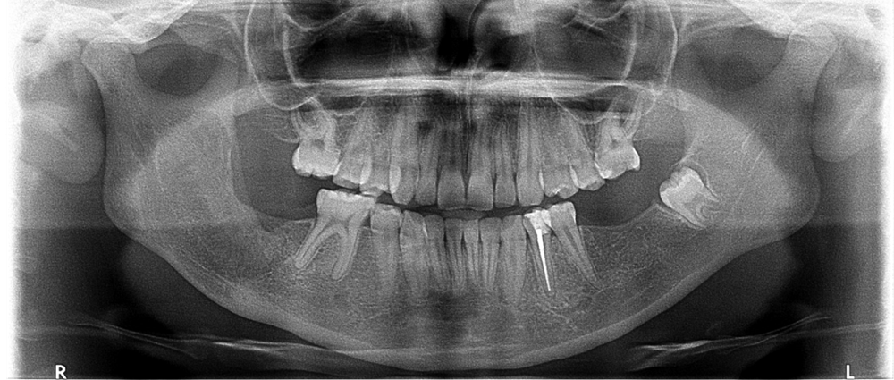
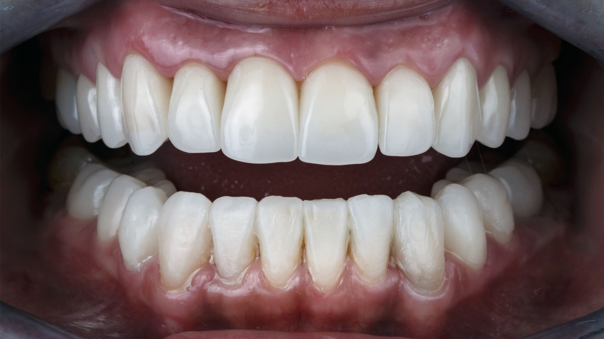
Odontogenic keratocyst is a different form of developmental odontogenic cyst that deserves special consideration because of its specific histopathological characteristics and clinical behavior. The ideal treatment for odontogenic keratocysts is a subject of debate among surgeons; however, treatments known as conservative among surgeons have shown good results when used as complementary treatment. Decompression treatment has been shown to be a promising alternative, but despite good results, this type of treatment depends of patient's cooperation. This paper aims to report a clinical case of an odontogenic keratocyst decompression in the right mandibular body and branch using Elixir Sanativo® Phytotherapy for irrigation. Then, enucleation and curettage were performed with Carnoy's solution. Significant remodeling of the cortical bone, extinguishment of the lesion and bone neoformation were observed, with no signs of relapse until now. There is a return of normal stomatognathic function in this clinical case report with a 2-year follow-up in total.
Keywords: Odontogenic Keratocyst, Decompression, Phytotherapy.
Expandir Resumo
Acessar Texto Completo
Ferimento provocado por arma branca impactada em região maxilofacial: Relato de caso
Resumo:
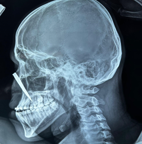
INTRODUCTION: Injuries to the maxillofacial region caused by stabbing weapons, despite being little reported, may present high risk for the patient. Moreover, because this area is composed of noble anatomical structures, penetrating injuries can be highly complex to treat due to their difficult access. CASE REPORT: Male patient 21 years old, victim of a stabbing injury, object stuck in the face, was attended to in the emergency department of the Sergipe Emergency Hospital, presenting on clinical examination with a penetrated foreign body in the back of nose region. After requesting an imaging exam the involvement of the affected area was confirmed with a subsequent surgical approach. DISCUSSION: Anatomical knowledge of the maxillofacial region is of paramount importance for better treatment management, since functional and aesthetic factors are involved in the resolution of these lesions.
Keywords: Stab wounds, wounds and injuries, maxillofacial trauma.
Expandir Resumo
Acessar Texto Completo
Contribuições do tratamento ortodôntico em pacientes com doenças periodontais
Resumo:
INTRODUCTION: The interaction between Orthodontics and Periodontics can contribute efficiently to dental treatment, being able to provide patients with periodontal disease, conditions so that periodontite health can restabilize and restore satisfactory occlusion. REVIEW: The present work proposed to evaluate, through literature review, such as articles, theses and books related to the subject, orthodontic treatment in patients with periodontal disease. DISCUSSION: From the information obtained it was possible to conclude that the orthodontic-periodontal union brings many benefits to the periodontium, by increasing the insertion capacity of periodontal ligament fibers to root surfaces, and thus establishing contact between the tooth and its respective alveolus. It should be emphasized that these benefits are considerable, provided that the biomechanical principles of orthodontics and the biological limitations of the periodontium are respected. FINAL CONSIDERATIONS: Finally, the present work showed that orthodontic treatment can be complementary to periodontal treatment, helping the patient to control biofilm and enabling a functionally and aesthetically stable occlusion.
Keywords: orthodontic treatment, periodontal treatment, ortho-perio union.
Expandir Resumo
Acessar Texto Completo
Panorama no ensino da Odontogeriatria na região sudeste do Brasil
Resumo:
Introduction: Odontogeriatrics started to be considered a specialty of dentistry from the year 2001, and Brazil was the first to recognize it. There are 244 specialists in Odontogeriatrics: 52 in Rio de Janeiro, 79 in São Paulo, 29 in Minas Gerais and 12 in Espírito Santo. Even with 19 years of existence, the number of professionals is reduced compared to the current elderly population. It is necessary that undergraduate students have basic knowledge about the particularities of the elderly, so that they can carry out individualized treatments and act in the prevention of possible damages. Objective: to carry out a survey of data in the faculties of Dentistry of the Southeast region of Brazil, recognized by the MEC, to verify how many present the discipline of Odontogeriatria, in a mandatory or optional way. Method: We analyzed the total number of Higher Education Institutions that presented: undergraduate dentistry, the discipline of Odontogeriatrics or related subjects, and make available the curriculum matrix. It was segregated between: offering the discipline (mandatory or optional) and those that did not provide the discipline. Results: Of the 161 colleges present in this region, it was found that 64% do not have the discipline of odontogeriatrics, 30.4% have the mandatory one, and 5.6% have it as an optional subject. Bearing in mind that there are 172 specialists in Odontogeriatrics throughout this region, and that only 36% of universities train professionals with knowledge of the particularities of dental care for elderly patients. Conclusion: There is a professional lack of preparation and an inability to provide quality care to the geriatric patient.
Keywords: Geriatric Dentistry, Dental Assistance for The Elderly, Dentistry, Seniors.
Expandir Resumo
Acessar Texto Completo
Fotografia digital na prática odontológica
Resumo:
Introduction: The use of digital photographs in dental clinics is increasing and allows the dentist a range of possibilities: it helps in the diagnosis and prognosis of dental diseases, in communication with colleagues and with dental laboratories, in addition, it is a supporting documentation judicial system and a means of publicizing and promoting the profession. Revision: Dental photography is an integral part of the dental record and communication with patients, so all patients should have photos taken of their initial and final condition. Almost 72% of dentists are adept at the technique, which is used mainly in orthodontics, aesthetic dentistry, in soft tissue analysis (nasopalatine cleft) and in the differentiation of pathologies in the oral mucosa (leukoplakia / lichen planus). Discussion: Knowledge of various methods of tampering and editing of images brings out the forgery. Digital photographs for dental records and legal protection must be standardized and closer to reality, that is, without modifications and effects that alter the original image. Final considerations: Digital photographs are often used for documentation, marketing, patient education, and peer-to-peer communication. However, the falsification of images for marketing purposes and the disapproval of some patients, due to the exposure of the image, discomfort and shyness in front of the cameras, prevent digital dental photographs from being used in all cases and mandatory recording in the medical record.
Palavras-chaves: Dental photography, digital photography, dentistry.
Expandir Resumo
Acessar Texto Completo
Osteonecrose maxilar induzida por agentes antirreabsortivos: conceitos, epidemiologia e conhecimento de profissionais e usuários
Resumo:
Introduction: Medications called antiresorptives can be used as adjuvants in the treatment of bone and/or bone cancer diseases, with bisphosphonates, RANK-L inhibitors and antiangiogenic agents as the main representatives of the class. Review: The three drug classes have in common the possibility of causing, from their use, a rare adverse effect, but with extreme morbidity called Drug-Induced Maxillary Osteonecrosis (ONMIM). Due to the increasingly widespread use of these drugs for the treatment of different tumor and bone diseases, the knowledge of the medical, dental and also lay people, representing the patients themselves, about the possibility of the occurrence of ONMIM has become increasingly important. Many patients using antiresorptive medications do not have the knowledge or were not instructed by their physicians about the risk of their adverse effects; which can lead to inadvertent dental treatment and the onset of the disease. Discussion: Prevention before the use of medications through a careful assessment of the oral health condition and an elaborate plan of care between the oncology team and dentists, can reduce the risk of developing ONMIM. Final considerations: Health professionals must take responsibility for sharing their knowledge with patients and emphasize the importance of prevention through dental follow-up. And so, knowledge and awareness can minimize the incidence of the disease through preventive care.
Keywords: bone density conservation agents, denosumab, osteonecrosis, bisphosphonate-associated osteonecrosis of the jaw, antirresorptive medication, bisphosphonates.
Expandir Resumo
Acessar Texto Completo
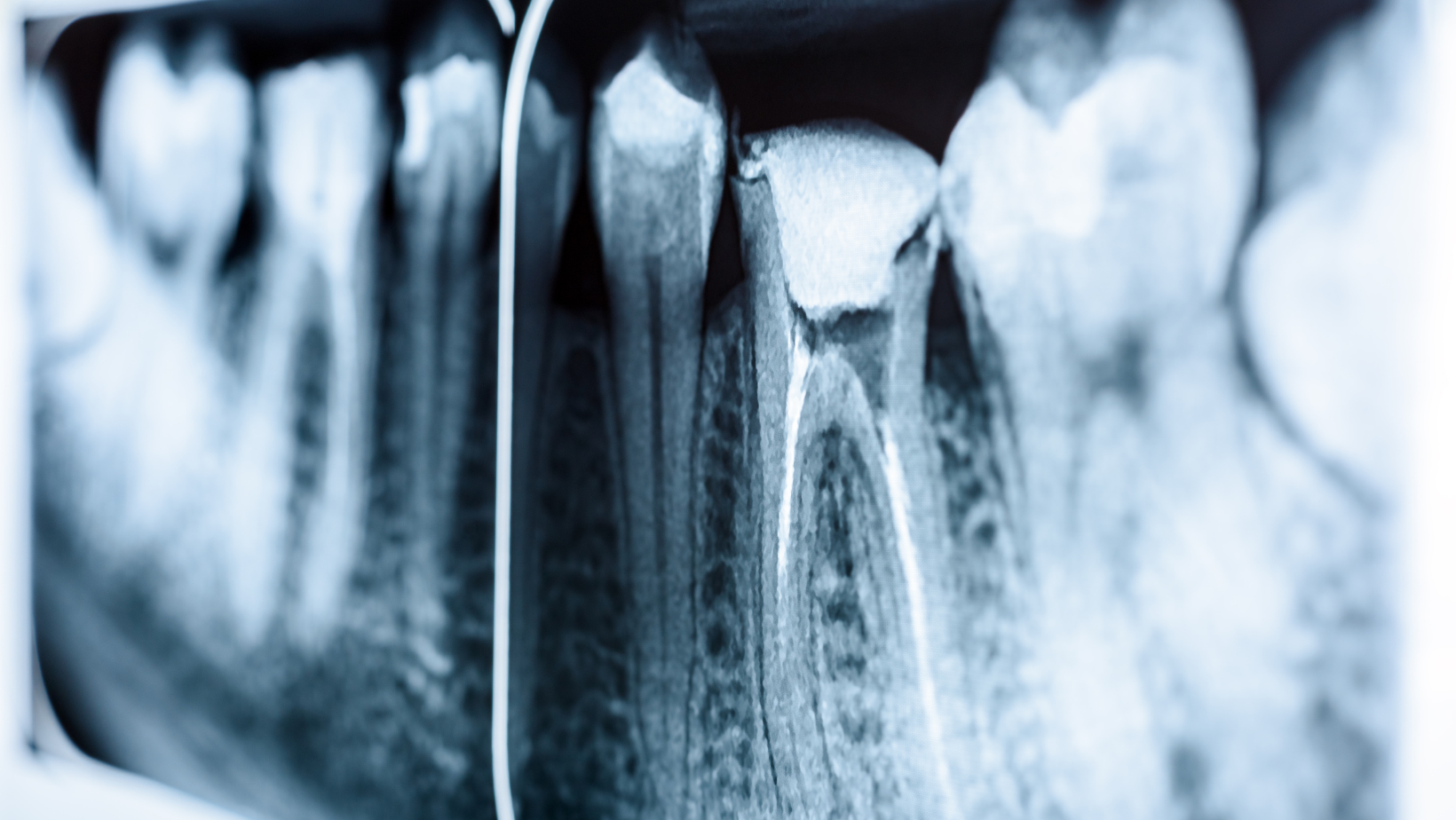
Tomografia computadorizada de feixe cônico: novos recursos e softwares para diagnóstico em endodontia
Resumo:
Abstract: Introduction: Cone-beam computed tomography (CBCT) is the most used in dentistry, due to its speed and low radiation compared to medical tomography; with this, it favored all specialties, especially endodontics. Review: To carry out this work, scientific articles, books, theses, dissertations and monographs related to the use of CBCT were researched, with emphasis on endodontics, especially in relation to current imaging resources. For this, online, national and international databases such as PUBMED, GOOGLE ACADÉMICO, SCIELO, Scopus and Web of Science were used. Discussion: Endodontics is one of the areas that most explores CBCT in dentistry. Visualization software/algorithms developers for image formation have developed resources for root canal measurement, file/instrumentation guides, making endodontic treatment safer and more predictable. For the endodontist, three-dimensional images help to clearly visualize the location of the apical foramen (which may vary), the size of the lesion that is in the periapical area, the presence of cracks/root fractures, whether root resorption can be resolved with treatment, among others. Other difficulties that periapical radiography cannot show, because it is two-dimensional. The transfer from analog to digital dentistry is still happening, prices are becoming affordable, ease and time saving is transforming and making developers have ideas for great innovations. Therefore, this study aimed to review the literature and discuss the new features of tomographic image viewer software. Final considerations: However, CBCT with three-dimensional imaging capability is of utmost importance for the endodontics of the future.
Keywords: endodontics, X-rays, software, cone-beam computed tomography.
Expandir Resumo
Acessar Texto Completo